
Total Body Photography (TBP) is used to assist with the diagnosis and monitoring of naevi (moles) in patients at higher risk of melanoma.
This includes patients with multiple naevi (many moles); a prior history of melanoma; a strong family history of melanoma; and/or a genetic susceptibility to melanoma.
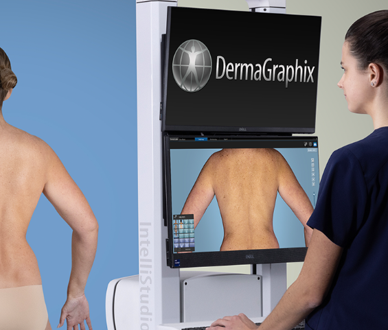
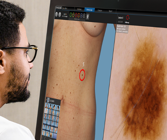
This advanced digital imaging technology provides precise documentation of naevi on a digital body map, allowing for enhanced monitoring of high risk patients. When TBP is repeated at chosen intervals by your dermatologist, it can be used to detect new and changing moles, with digital side-by-side comparison. Sequential Digital Dermoscopy Imaging (SDDI) or close up individual mole imaging, is also used alongside TBP for enhanced monitoring of selected lesions. TBP and SDDI have integrated Artificial Intelligence for annual automated total body comparison and short term targeted mole comparison. Currently this AI is not clinically validated and is not used for clinical purposes. All imaging is supervised by your dermatologist and all decisions are made/shared between patient and dermatologist.
TBP and SDDI may assist with earlier detection of melanoma and reduce benign excisions in this higher risk population. TBP is provided onsite at our facility and is performed by our highly trained registered nurses/melanographers, who have undergone additional training in skin imaging. TBP is an adjunct to your routine surveillance full skin examination with your dermatologist.
Please note TBP + SDDI may take 15 min – 1 hour. All patients are required to see our dermatologists prior to assess for suitability. All make-up and nail polish should be removed prior to imaging. Hair should be tied back and if there is significant body hair you may be asked to clipper this prior to attending for imaging.
Referrals for enhanced surveillance with total body photography from general practitioners, skin cancer clinics, dermatologists, oncologists and surgeons are welcome.